How the iTero Lumina Improves Digital Dental Scans in Orthodontics.

Dr. David Boschken is the owner of Boschken Orthodontics in the San Francisco Bay Area.
He is the longest-tenured faculty member and Invisalign lecturer, preparing clinicians worldwide for over 25 years. He received his dental degree and training in orthodontics from the University of Pennsylvania’s School of Dental Medicine.
Limitations experienced by orthodontic teams and patients.
Digital orthodontic practices faced several limitations in the past during new patient consults and follow-up visits. Some of the common obstacles my team and I encountered with previous iTero™ scanner models were the size and weight of the wand, the need for a developed technique to capture clear scans in a reasonable amount of time, the learning curve for new staff to become proficient in scanning, and the challenges of obtaining scans from hard-to-reach areas like the distal surfaces of terminal teeth, deep palates, interproximal surfaces, and undercuts, among others.
From the patient’s perspective, we noticed some patients experienced discomfort during the scanning process caused by the wand’s size and the scan’s duration. These situations were especially evident for patients with temporomandibular discomfort, small mouths, special needs such as those with craniofacial malformations as well as persons who have dental phobia. However, it is important to note that these discomforts are not as prevalent or severe as they were with traditional impressions, which we last used over a decade ago.
To improve the scanning experience for clinicians and patients, and to enhance the performance from speed to quality of visualizations and clinical outcomes, Align Technology looked for solutions in innovative design and technology.
iTero Lumina™ scanners.
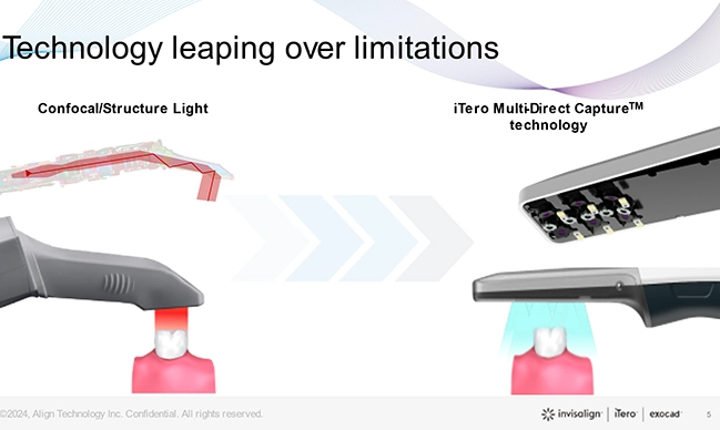
Comparison between today’s wands size and technology and the iTero Lumina™ scanners with the iTero Multi-Direct Capture™ technology.
Align Technology listened to orthodontic teams and patients and developed an innovative technology to overcome those limitations. They realized that building upon the confocal technology to improve those obstacles was not feasible.
Previous iTero™ scanners and most scanners on the market have a single camera in the base of the wand that indirectly captures one to two teeth at a given time. This concept is defined as the field of capture, and operators would prefer a scanner with a larger field of capture to include more teeth and tissues at any given time without compromising image quality. However, companies would have to design larger wands to achieve a larger field of capture with the known technology, which is inconvenient for clinicians and patients alike. The ideal scenario is having a small and lightweight wand with a larger field of capture that can scan at the operator’s preferred speed rather than the scanner dictating the speed while maintaining high-quality images.
The most apparent improvement in the new scanner is the wand size and its weight. The iTero LuminaTM intraoral scanner wand is 50% smaller and 45% lighter than previous models1. This unit also offers 3x larger field of view designed for more surface area capture enabling faster scanning2. Inside the wand, developers introduced the iTero Multi-Direct Capture™ technology (MDC) that allows for simultaneous multi-angle capture that reduces maneuvering3 and allows for a smaller wand. But this scanner also has six cameras - five additional than previous models - that capture details as small as 30 um, resulting in high quality photos4.
Using a smaller and lighter wand for orthodontic scanning impacts practice workflows. Today, some practices have chosen to scan patients only after accepting orthodontic treatment as they have yet to discover the value of adding a few extra minutes to the initial consult. However, this approach can be risky as it may prevent prospects from fully understanding their oral health needs, and they may walk away without accepting treatment. Unfortunately, such practices are missing out on other valuable features of iTero scanners, such as the iTero Occlusogram and the Invisalign® Outcome Simulator Pro with in-face visualization. These tools aid in education and communication, especially during new patient visits. New clinicians rely on these tools to enhance communication during new patient visits to help increase treatment acceptance. Failure to scan every individual in your office could result in fewer starts and suboptimal production5.
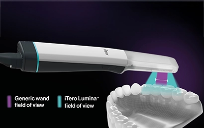
The wide field of capture results in capturing larger teeth segments at each moment during the scan, reducing scanning time6 and improving accuracy7.
High-accuracy full arch scan and effortless scanning promote a short learning curve and increased utilization9.
However, with the iTero Lumina™ scanner, you can scan two times faster compared to previous iTero™ intraoral scanners10 , and inexperienced practices will not have to be concerned about added time to complete a scan on potential patients. The onboarding with this scanner was easy. The new effortless scanning software has vastly improved the way we scan patients. The previous technique of scanning on one quadrant and then “rolling” around without lifting the wand is no longer necessary. With iTero Lumina™, the assistant can scan, pick up, and move to a different location inside the mouth without interrupting the results. With the improved depth of scans11 and three times field of view6, we saw a considerable improvement in scanning times, quality of data captured, and hence the quality of aligner fit.
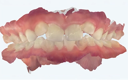
The photorealistic 3D model from the iTero Lumina™ (left) compared to the 3D model obtained with an iTero Element™ 5D Plus series scanner (right).
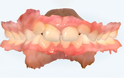
In addition to being easy to use, fast, and comfortable for both the operator and the patient, the iTero Lumina™ scanner impressed me with the quality of the 3D models and intraoral photos it generated. This device does not need to touch the teeth or soft tissues like previous models. The iTero Lumina™ scanner can capture depths of 25mm11, which is crucial when scanning patients with deep12, narrow palates12, or acute gag reflexes. The wider field of view facilitates an easier scan of the palate with no added time or effort, almost as a by-product of the dentition scan.
Moreover, the addition of multiple angled cameras creates accurate and natural 3D color models and dimensional images. The high resolution and detail captured by these cameras are comparable to the renderings produced by digital single-lens reflex (DSLR) cameras13, the standard in orthodontics and dentistry. These enhancements have significantly contributed to building trust and improving communication and engagement during consultations with new patients. Prospective patients can now see what I see inside their mouths on the scanner screen during an intraoral exam. They can easily follow my explanations, which have become shorter since the visual representation is right before them.
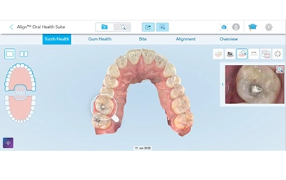
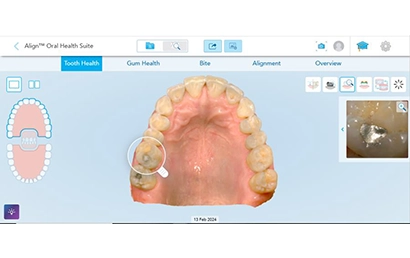
As a result of the high-quality scans and intraoral images, I eliminated images from digital cameras for Invisalign® cases15. My practices solely rely on the scan to produce intraoral photos that are saved in the cloud and easily accessible on the myiTero.com portal and my practice management software. This new workflow has helped us save valuable clinical and administrative time while collecting and storing high-quality records efficiently and effectively.
The photorealistic 3D model13 and the intraoral photos from the iTero Lumina™ (left) compared to the 3D model obtained with an iTero Element™ 5D Plus series scanner (right). The effortless scanning16 and the increased field of capture6 allow operators to obtain more data in fewer seconds.
In the past 12 months of assessing this scanner, we have seen a measurable increase in case acceptance. Our practice data has shown scanning quality and times. During the consultation, the iTero Lumina™ scanner has given us the edge over our competition. Combining the Invisalign® Outcome Simulator Pro simulation to generate real-time before and after images of their smile gives the prospective patient all the necessary visuals to make that final decision to start treatment. Just showing potential patients the six cameras and viewing the lifelike scanning images on the screen often is enough to close same-day starts.
Dr. David R. Boschken has been providing Invisalign® aligner treatment to patients in the San Francisco Bay Area since 2000, and he has successfully treated over 6,000 patients. He founded several clear aligner treatment planning companies and consulting services. Dr. Boschken is also the longest-tenured faculty member and Invisalign lecturer, preparing clinicians worldwide for over 24 years. He graduated from the University of California, Berkeley, with a double major in Biochemistry and Anthropology. He received his dental degree and training in orthodontics from the University of Pennsylvania’s School of Dental Medicine. After graduation, Dr. Boschken completed a hospital residency at Guy’s and St. Thomas Hospital in England. In addition to owning two private practices in Los Altos and San Jose, California, Dr. Boschken enjoys boating and skiing in Tahoe with his family during his free time.
- The iTero Lumina™ intraoral scanner wand is 50% smaller and 45% lighter than the iTero Element™ 5D imaging system wand. Compared to iTero Element™ 5D imaging system wand, excluding the wand cable. Data on file at Align Technology, as of November 15, 2023
- Compared to the field of view of the iTero ElementTM 5D imaging system, when the iTero LuminaTM intraoral scanner’s scanning distance is 12 mm. Data on file at Align Technology, as of November 15, 2023.
- The iTero Lumina™ intraoral scanner is powered by the new, proprietary iTero Multi-Direct Capture™ technology. The iTero Lumina™ intraoral scanner offers simultaneous multi-angle capture designed for less maneuvering. Data on file at Align Technology, as of November 15, 2023.
- The iTero Lumina™ intraoral scanner features integrated high resolution intraoral cameras for capturing details as small as 30 um. Data on file at Align Technology, as of November 15, 2023.
- Scanning decision at independent discretion of health care provider.
- The iTero LuminaTM intraoral scanner offers 3x larger field of view designed for more surface area capture enabling faster scanning. Compared to the field of view of the iTero ElementTM 5D imaging system, when the iTero LuminaTM intraoral scanner’s scanning distance is 12 mm. Data on file at Align Technology, as of November 15, 2023.
- The iTero LuminaTM intraoral scanner has scientifically proven greater accuracy for your clinical orthodontic needs. Greater global accuracy in comparison with the global accuracy of the iTero ElementTM 5D imaging system. Data on file at Align Technology, as of November 15, 2023.
- iTero LuminaTM demonstrated superior full jaw accuracy compared to tested competitors. Based on bench testing conducted using ADA/ANSI 132 standard Model simulating “long distance accuracy (full jaw measurement)” in July 2024. Sample definition: 3 operators performed 30 repetitive scans with each tested scanner; Sample size: n=90 (3x30) scans for each scanner tested; Tested scanners: iTero LuminaTM, Trios 5, CS3800, Medit i700, Alliedstar; Results: The accuracy of iTero LuminaTM is significantly higher than that of all 4 competitors; with a reduction of total error ranging from 0.11% to 0.46%. Accuracy was defined as the accumulated average error + STD of all measurements specified in the standard. Data on File at Align Technology, as of August 26, 2024.
- 81% of surveyed users agree that the iTero LuminaTM intraoral scanner scanning experience helps remove adoption and utilization barriers. Based on a survey in September 2023 of n=22 users who participated in a global limited market release, working with iTero LuminaTM intraoral scanner for an average period of 6 months, representing both Invisalign® trained general practitioners and orthodontists, and their staff in NA, EMEA and APAC, who were presented with a 4 point level of agreement scale from strongly agree to strongly disagree with the following statement: “The iTero LuminaTM intraoral scanner scanning experience helps remove adoption and utilization barriers.” Data on file at Align Technology, as of November 15, 2023.
- The iTero Lumina™ intraoral scanner is designed to enable 2x faster scanning compared with previous iTero™ intraoral scanners. Compared to the iTero Element™ 5D imaging system with tolerance AVE=±0.1 operating at a working distance from 0-20 mm. Data on file at Align Technology, as of November 15, 2023.
- The iTero Lumina™ intraoral scanner boasts a 25 mm capture distance. Data on file at Align Technology, as of November 15, 2023.
- The iTero Lumina™ intraoral scanner’s Multi-Direct Capture™ technology, with its wider field of view1, large capture distance2 and multi-angle capture3, is designed to simplify capture of challenging areas like the palate, edentulous spaces, partially erupted teeth and crowded teeth.
- The iTero Lumina™ intraoral scanner offers 3x larger field of view designed for more surface area capture enabling faster scanning. *Compared to the field of view of the iTero Element™ 5D imaging system, when the iTero Lumina™ intraoral scanner’s scanning distance is 12 mm.
- The iTero Lumina™ intraoral scanner boasts a 25 mm capture distance.
- The iTero Lumina™ intraoral scanner offers simultaneous multi-angle capture designed for less maneuvering. Data on file at Align Technology, as of November 15, 2023
- Majority of surveyed users agree that the iTero Lumina™ intraoral scanner’s 3D model is comparable to that of an intraoral photo. For Invisalign® record-taking cases only. Based on a survey in September 2023 of n=22 users who participated in a global limited market release, working with iTero Lumina™ intraoral scanner for an average period of 6 months, representing both Invisalign® trained general practitioners and orthodontists, and their staff in NA, EMEA and APAC, who were presented with a 4 point level of agreement scale from strongly agree to strongly disagree with the following statement: “The iTero Lumina™ intraoral scanner 3D model is comparable to that of an intraoral photo.”
- Majority of surveyed users agree that the iTero Lumina™ intraoral scanner’s superior 3D model boosts patient engagement. Based on a survey in September 2023 of n=22 users who participated in a global limited market release, working with iTero Lumina™ intraoral scanner for an average period of 6 months, representing both Invisalign® trained general practitioners and orthodontists, and their staff in NA, EMEA and APAC, who were presented with a 4 point level of agreement scale from strongly agree to strongly disagree with the following statement: “The iTero Lumina™ intraoral scanner’s superior 3D model boosts patient engagement.” Data on file at Align Technology, as of November 15, 2023
- iTero Lumina photorealistic scans are clinically comparable to intra-oral photography. Based on clinical data collected in January 2024 of n=15 subjects who participated in a clinical study with iTero Lumina™ intraoral scanner. Data reviewed by Invisalign trained practitioners (Orthodontists and GPs) in conjunction with Align clinical team. Data on file at Align Technology, as of January 31, 2024.
- 90% of surveyed doctors and their staff agree that they feel confident when scanning with the iTero LuminaTM intraoral scanner due to its reliable and effortless scanning experience. Based on a survey in September 2023 of n=22 users who participated in a global limited market release, working with iTero LuminaTM intraoral scanner for an average period of 6 months, representing both Invisalign® trained general practitioners and orthodontists, and their staff in NA, EMEA and APAC, who were presented with a 4 point level of agreement scale from strongly agree to strongly disagree with the following statement: “I feel confident when scanning with the iTero LuminaTM intraoral scanner due to its reliable and effortless scanning experience.” Data on file at Align Technology, as of November 15, 2023.