Efficient end-to-end digital dentistry workfows in a multidisciplinary practice - Same-day 3D-printed resin crowns for a pediatric patient.
Meeting the needs of the modern dental patient
Today’s patients are more informed and demanding than ever before. With a wealth of information readily accessible online, families seek dental care that not only addresses their children’s clinical needs but also aligns with their expectations of efficiency, comfort, and transparency. Young patients often have limited attention spans and may experience anxiety in the dental chair, making time-effective treatments crucial. Simultaneously, dental practices strive to maintain profitability amid varying economic conditions while providing high-quality care.
Parents increasingly question professional advice; the 2024 Edelman Trust Barometer survey1 indicates that distrust in healthcare providers is trending upwards. Approximately 58% of respondents reported that contradictory expert advice, changing health recommendations, and a lack of clear information prevented them from taking better care of their family’s health.
Clear communication becomes essential to bridge this trust gap.
By leveraging technological aids, we can enhance transparency and understanding for both children and their parents. These tools help families visualize and understand complex information and build confidence in our diagnoses and treatment plans.
As dentists, we have the opportunity to transform the way we practice to meet these evolving needs. Embracing advanced digital solutions is critical to enhancing the treatment experience for child patients, building trust with parents, and improving the efficiency of our practices.
The importance of an integrated digital ecosystem in dentistry
Traditionally, dental practices have relied on analog solutions and fragmented pieces of technology operating independently, leading to inefficiencies and potential errors. For example, separate systems for patient management, treatment planning, CAD design, and laboratory communication often require manual data transfers and lack seamless integration. This fragmentation consumes valuable time and makes it challenging for practitioners and staff to fully leverage the capabilities of digital dentistry.
In contrast, an integrated digital ecosystem like the Align™ Digital Platform connects (Figure 1) many aspects of dental care— data acquisition, diagnosis, patient communication and conversion, treatment planning, execution, and collaboration—within a unified platform. It offers an end-to-end digital treatment experience, generating interconnected workflows and treatment solutions – the Align Digital Workflow – supporting you from the first consultation through to the final smile.
One of the innovations in this workflow is the iTero™ Design Suite. Using the power of exocadTM CAD/CAM software, it offers simplified doctor and staff-friendly applications to design for in-practice 3D printing of models, bite splints, restorations and mock-ups.
The iTero Design Suite™ is designed to shorten time to treatment and provide high-quality solutions within the practice for improved patient experience.
This paper outlines our approach to treating child patients following the Align™ Digital Workflow, implemented at the Artist Dental Clinic. We aim to showcase through practical insights how this workflow helps us to meet patients’ expectations and achieve superior clinical results.
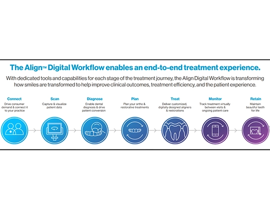
Figure 1
Figure 1: Align’s technological solutions offer end-to-end digital workflows for a multidisciplinary practice.
Case Study: Same-day 3D-printed resin crowns for a pediatric patient
Patient presentation
A few months ago, the parents of a young girl brought her to our clinic for a consultation. We obtained diagnostic radiographs and performed an intraoral scan using our iTero™ 5D Plus imaging system during the examination. The patient exhibited poor oral hygiene and had multiple carious lesions on teeth 54, 55, 64, 65, 75, 84, and 84(Figure 2). Additionally, the lower central incisors were found to be congenitally missing, necessitating future orthodontic intervention as she matures.
Digital aid for diagnosis and enhanced communication
The pediatric dental treatment often includes challenges. Among them, the effective communication with parents and establishing an agreement with the child to ensure compliance. Clear communication with parents is essential for providing high-quality care, as they need to understand their child’s oral health condition and the required treatment to give informed consent. Before acquiring an intraoral scanner, I used either a dental mirror or a digital camera to discuss oral health issues with parents. However, using a dental mirror proved difficult, as parents found it challenging to see into the small mirror—especially given the child’s tiny mouth and teeth. While taking intraoral photographs offered better visualization, it necessitated the use of retractors and mirrors, which could cause discomfort to the child. This discomfort sometimes compromised further treatment by disrupting the initial connection established with the child.
Figure 2 a-d: A comprehensive digital suite for oral health consultations, Align™ Oral Health Suite, enables us to visualize diagnostic findings using intraoral camera images and iTero™ NIRI (Near Infra-Red Imaging). Tooth 54 (a), tooth 52 (b), tooth 72 (d), tooth 84 (c).
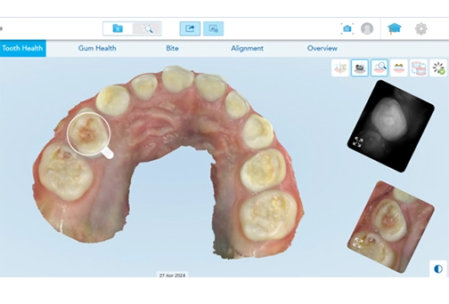
Figure 2a
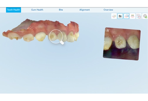
Figure 2b
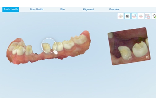
Figure 2c
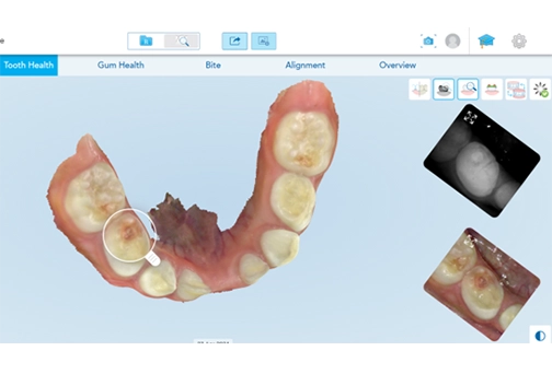
Figure 2d
The iTero™ intraoral scanner helps me address both challenges simultaneously. In my experience, performing an intraoral scan is more manageable than capturing intraoral photographs in children. I usually begin by scanning a small area and then direct the child’s attention to the screen. Children enjoy watching the dynamic images, becoming fully engaged with the “movie” on the screen, which helps them remain calm.
When we communicate with parents, reviewing the scans using the Align™ Oral Health Suite on a large screen helps them to quickly comprehend the extent of their child’s dental issues and the necessary interventions. The photorealistic 3D models and high-definition intraoral images captured with the iTero™ intraoral scanner facilitate clear visualization of patient’s dentition and the extent of decay. In my experience, when parents gain a clear understanding of child’s oral health conditions, they are more likely to trust the proposed treatment and to provide informed consent.
Treatment plan
Considering the family’s residence far from the clinic, the extensive treatment required, and the patient’s age, we decided to perform the treatment (Figure 3) in a single visit under sedation. This approach minimized the number of appointments and reduced stress for both the patient and the parents.
Carious lesions on teeth upper right primary second molar (Tooth 55), upper left primary first molar (Tooth 64), upper left primary second molar (Tooth 65), lower left primary second molar (Tooth 75), and lower right primary first molar (Tooth 84) were restored using direct composite. The upper right primary first molar (Tooth 54) presented an advanced lesion with pulp exposure, necessitating endodontic treatment, a core build-up(Figure 4), and a crown restoration to restore its stability and function. Similarly, tooth 84 had an extensive carious lesion, so a crown restoration was necessary.
Teeth 54 and 84 were prepared (Figure 4, 5), and the preparations were scanned using the iTero™ 5D Plus imaging system. For this case, we’ve placed the margin line approximately 0.5 mm above the gingiva. Supragingival margins allow for an easier scanning process and favor superior periodontal health.
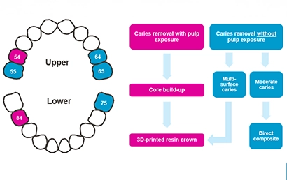
Figure 3: The decision flow for addressing patient’s needs.
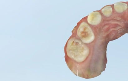
Figure 4a: Tooth 54 – Initial situation
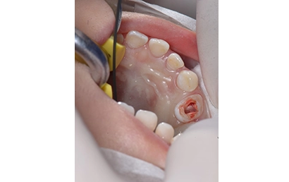
Figure 4b: endodontic treatment
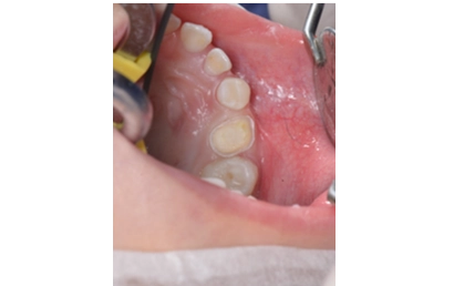
Figure 4c: core build-up
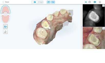
Figure 4d: final preparation scan
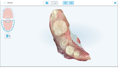
Figure 5: Tooth 84 – preparation scan
Treatment process for the 3D printed resin crowns
While I continued working on the direct restorations, a staff member designed the crowns using the iTero™ Design Suite. This CAD software integrates with the MyiTero.com cloud platform and can be launched directly from the patient’s file, automatically importing their data to start the design process immediately. The software features an automated wizard-guided workflow that leads the user through each step to complete the designs.
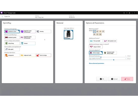
Figure 6: Restorative settings screen of the iTero™ Design Suite
The design process starts by verifying the type of restoration to be designed and production settings. This step is optional and could be skipped if the settings were predefined earlier (Figure 6).
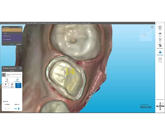
Figure 7: Margin line verification
Next is the restoration design.
We then review the auto-detected margin line and adjust it if necessary. In our experience, margin line tracing is usually accurate and doesn’t require adjustments often. (Figure 7).
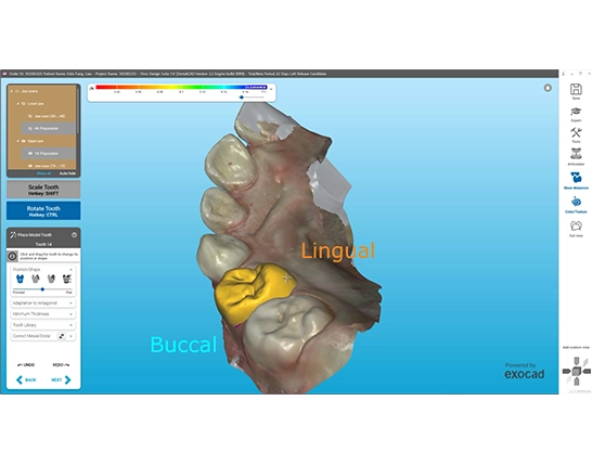
Figure 8: Designing the restorations
After confirming the margins and insertion direction, we adjust the shape of the crown, including the proximal and occlusal contacts (Figure 8).
Once the design is completed and reviewed, we transfer the files to the supported 3D printer software with a single click. (Figure 9 a-b).
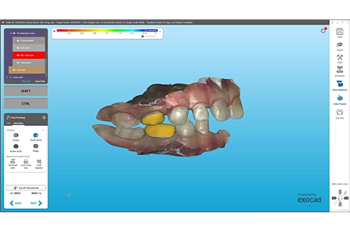
Figure 9a: Final review of the restorations on occlusion
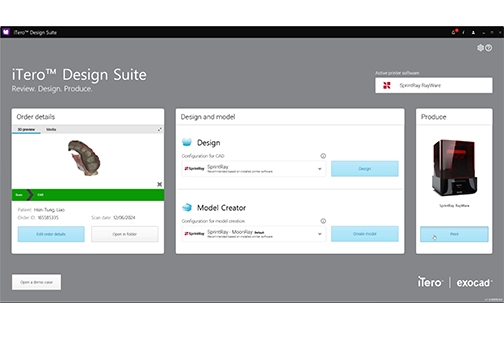
Figure 9b: Completed design files transferred to the 3D printing software
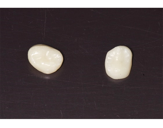
Figure 10: 3D-printed crowns prepared for cementation
We fabricated the crowns in-house using our 3D printer and ceramic resin. The design process took about five minutes, printing approximately eight minutes, and washing and curing another five minutes. From taking the scans to having the crowns ready for cementation(Figure 10, the entire process took less than 20 minutes.
After cementation (Figure 11), the occlusion was optimal, and no occlusal adjustments were needed.
Figure 11 a-c: Long-term 3D printed resin crowns.
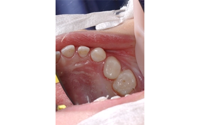
Figure 11a: Tooth 54
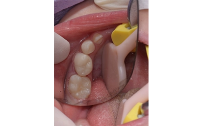
Figure 11b: Tooth 84
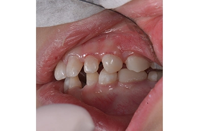
Figure 11c: Teeth 54 and 84 in occlusion
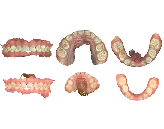
Figure 12: Intraoral scan images compare the initial situation (top) with the current patient condition (bottom) after the treatment
Post-treatment monitoring
About a month after the treatment, we scheduled the patient for a follow-up appointment to monitor treatment stability and the patient’s oral health status. We performed a scan with our new iTero™ Lumina scanner and compared the difference with the initial situation (Figure 12).
The new iTero Lumina™ intraoral scanner offers a field of view three times larger than that of the iTero Element™ 5D imaging system2. This enhanced view allows us to capture more surfaces at once and complete the scanning process much faster. As a result, we find that patients benefit from a more streamlined experience, and we have a more efficient workflow.
Due to the patient’s young age, the congenitally missing lower central incisors do not require immediate intervention. We will continue to monitor the situation as part of the patient’s ongoing dental care. The iTero™ scanner’s ability to store patient records and facilitate precise comparisons over time allows us to track developments in the patient’s dentition closely. This capacity for monitoring is invaluable in detecting any changes early and planning future orthodontic, restorative, or preventive interventions if necessary.
Conducting a post-treatment appointment allowed us to confirm the positive clinical outcomes of our treatment and demonstrate to the parents the improvements in their child’s oral health resulting from the quality care we provided. This approach helps us build strong relationships with patients and turns them into loyal advocates for our practice.

Dr. Yi Jyun Chen is a specialist orthodontist with extensive expertise in aesthetic dentistry.
He earned his Bachelor of Dental Surgery with Special Contribution Honors and a Master of Dental Science from Chung Shan Medical University, where he also completed his postgraduate training in orthodontics. In 2012, he obtained his doctorate, focusing on laser applications in orthodontics.
Dr. Chen lectures extensively on Aesthetic Dentistry and Orthodontics and is an Align Faculty member. Since 2017, he has been practicing privately at Artist Dental Clinic in Taichung City, Taiwan.
- 2024 Edelman Trust Barometer. (n.d.). Edelman. https://www.edelman.com/trust/2024/trust-barometer.
- The iTero Lumina™ intraoral scanner offers 3x larger field of view designed for more surface area capture enabling faster scanning. *
*Compared to the field of view of the iTero Element™ 5D imaging system, when the iTero Lumina™ intraoral scanner’s scanning distance is 12 mm. Data on file at Align Technology, as of November 15, 2023
The opinions expressed in this publication are those of the author and may not reflect those of Align Technology, Inc. The authors were paid an honorarium by Align Technology, Inc. in connection with this publication.