The Align™ Oral Health Suite, the latest tool on the iTero Element™ Plus Series imaging system.
Building trust and enhancing communication during oral health exams.
Author:
Dr. Joshua Austin, restorative dentist
San Antonio, TX
When meeting with a prospective patient for the first time, building trust can be a challenge in a dental clinical environment.
At our clinic, we aim to understand the individual’s needs, provide the best solutions, and establish a long-term doctor-patient relationship. Traditionally, we gather their dental and medical history, take records such as radiographs, conduct a clinical visual exam, and use the iTero Element™ 5D Plus imaging system to scan the patient.
The iTero™ intraoral scanner enables us to create a digital STL file with a 3D model of the person’s oral cavity in just a few minutes. This digital model offers us access to various tools to illustrate clinical findings. The Align™ Oral Health Suite is the integration of multiple proprietary iTero™ visualization and diagnostic aid tools into one easily accessible interface.
With the Align™ Oral Health Suite, we can demonstrate within minutes what we see inside the patient’s mouth from different perspectives. This allows us to communicate and educate new patients effectively. They can now see their mouths from various angles, and I can compare scans from two different time points to understand how specific interventions have helped improve their oral health over time.
In our experience, the Align™ Oral Health Suite helps us build trust within minutes and make consultations more meaningful and actionable, often leading patients to accept recommended treatment on day one. Finally, we conclude our consultations by sharing the iTero™ scan report, which summarizes the clinical findings in a composite of images captured by the snapshot feature. It includes notes highlighting the areas that require attention, enabling patients to make informed treatment decisions. By incorporating the Align™ Oral Health Suite, we are confident in our ability to provide optimal solutions while establishing a long-term doctor-patient relationship.
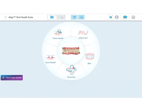
Figure 1
Case information
After being absent for over a year, the patient presented for a periodic oral health exam. We had a previous scan from him, which we used to compare to the current oral health conditions using the Align™ Oral Health Suite. After scanning the patient’s oral cavity and submitting the scan, we accessed the tool, which presents five conditions in the shape of a wheel. (Figure 1).
Figure 1: The landing page displays a wheel with five conditions, and the clinician can select where to start the consultation based on the patient’s needs observed and the chief complaint.
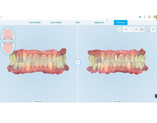
Figure 2
Overview
We started showing the Overview condition to provide a general panorama of the oral health status. Using the side-by-side 3D compare feature we discussed general changes that occurred in the past year. (Figure 2).
Figure 2: Within the Overview condition, I used the 3D side-by-side compare tool to contrast this patient’s oral health condition in April 2021 with his condition in May 2023. In 13 months, this patient’s oral health condition declined substantially. Overall, the following findings facilitated the conversation with the patient.
- White spots progressed into cavitated lesions that require composites, clear evidence helped the patient understand and acknowledge the need for treatment.
- The oral hygiene did not improve, with evidence of dental plaque, impacted food, and calculus, especially in the lingual areas of mandibular canines and incisors, helping him understand why we need to start this phase with a deep cleaning, and a prescription of fluoridated toothpaste to prevent incipient caries from progressing to cavitated lesions. Communicating the findings is only one part of an effective consultation. We also explain the consequences of not accepting the optimal treatment, while also offering treatment alternatives.
One of the advantages of the Align™ Oral Health Suite is that it provides the structure to conduct your consultations. Following this structure, we proceeded to review other conditions.
Tooth Health
Under the condition Tooth Health, the iTero™ NIRI technology (Near Infra-Red Imaging), the integrated 3D intraoral camera, and the stone model are the tools available to assess in detail the health of hard tissues and the conditions of restorations.
When we showed the patient the findings, he reached out to touch the screen to enlarge and move the 3D digital model around. This broke the ice and made him participate in the discovery process. Generally, I replicate what I see during my visual clinical exam in the imaging system’s screen. The difference is that when I do it in the patients’ mouths, they cannot see it, nor can they participate in the process. In a way, this is how my exams differ, because technology allows the patient to participate actively. Not only do the visualizations help me communicate and educate them, but also the interactions with the visuals from the patient make them more engaged in the process, helping me build that trust within minutes.
Leveraging the 3D model and the integrated 3D intraoral camera, we showed the patient how the white spot lesions from 13 months ago became cavitated lesions in teeth number 8, 9, and 10 (upper central incisors and upper left lateral incisor). Furthermore, the white spot lesions in the remaining upper and lower incisors and the canines expanded. A few teeth also revealed crazed lines and new white spots in the enamel. (Figure 3).
The crown in tooth number 3 (upper first right molar) presents damage in the porcelain near the gingival line on the palatal surface (Figure 4), and the crown on tooth number 15 (upper second left molar) in the palatal surface presents with a significant gingival recession with the presence of debris and soft tissue when probed with an explorer (Figure 5). Significant areas of recessions are present on the buccal surface of tooth number 3 (upper first right molar), and on the buccal surfaces of the upper left premolars (teeth number 12, 13).
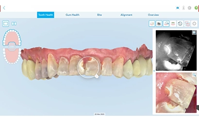
Figure 3: Incipient caries progressed into cavitated lesions in the upper incisors. Closeups of individual teeth are available through the iTero™ NIRI technology ((Near Infra-Red Imaging) tool and the integrated 3D intraoral camera.
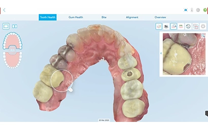
Figure 4: The integrated 3D intraoral camera supports the assessment of hard and soft tissues and the condition of restorations. The crown on tooth number 3 (upper first right molar) presents damage in the porcelain near the gingival line in the palatal surface.
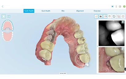
Figure 5: The integrated 3D intraoral camera assists in detecting a considerable gingival recession with potential radicular caries in the palatal surface near the gingival margin of tooth number 15 (upper second left molar). Tooth number 13 (upper second left molar presents an occlusal restoration to seal the access when an endodontic treatment was previously completed.
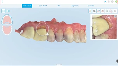
Figure 6: Highlights a substantial gingival recession exposing the root in the buccal surface of the upper right first molar.
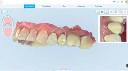
Figure 7: Shows gingival recession on the buccal surface of the upper left premolars. The integrated 3D intraoral camera facilitates communication and education during the consultation.
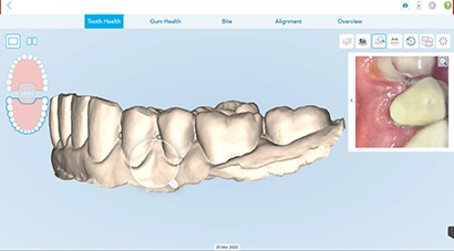
Figure 8: The stone model view aids in visualizing tooth wear, such as gingival recession, erosion, attrition, and abfraction. The lower left premolars show significant recession of the gingiva, exposing the roots and the cervical margin of the restorations.
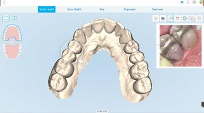
Figure 9: Illustrates wear in the amalgams in the upper right premolars and the upper right central incisor and canines.
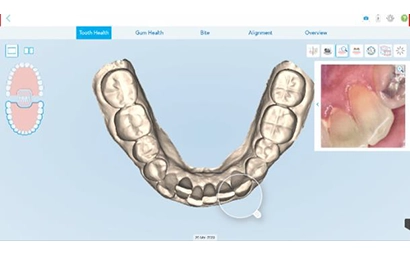
Figure 10: Highlights tooth wear across the incisal edges of canines and incisors and buccal cusps of the premolars.
In the lower arch, both left premolars showed significant recessions in the buccal surfaces. When switching to the stone model mode, these recessions became more evident, and we also found wear in the enamel and restorations (Figure 8). In the upper arch, we identified wear in the amalgams in the upper right premolars and in the incisal edges of the canines, lateral incisors, and the right central incisor. In the lower arch, we identified wear in the incisal edges of all anterior teeth and in the buccal cusps of the premolars (Figures 9-10).
Gum Health
The Gum Health condition activates the stone model mode and the integrated 3D intraoral camera. The latter aids us in assessing the color, volume, and texture of the soft tissues and the presence of dental plaque, calculus, and impacted food. Looking at the 3D model in stone mode helped us evaluate for gingival recession, abfractions, attrition, and erosion. We had looked at these under the Tooth Health condition; however, here we can view it from the soft tissue aspect. Moreover, using iTero™ TimeLapse allowed us to compare the current scan with a previous one and highlight changes in the gingival margins. Teeth 20 and 21 (lower left premolars) experienced significant recession in just 13 months. Furthermore, we also discovered crowns with compromised fit that progressed into dental and radicular caries or teeth with exposed root cementum, increasing the risk for sensitivity or further carious lesions in the future.
Finally, we discovered calculus deposits in the lingual gingival area of lower incisors and canines. The sideby-side 3D compare tool was useful for this case to illustrate the recurrent issue with oral hygiene (Figure 11).
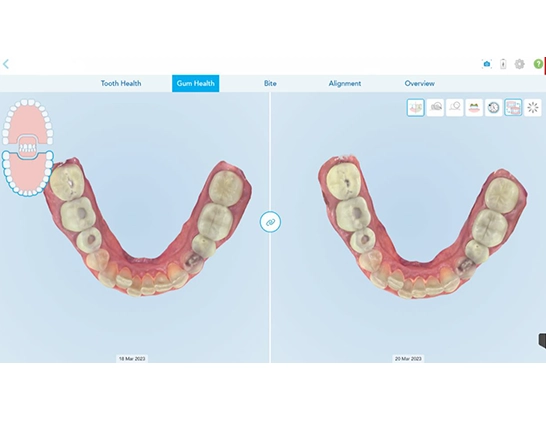
Figure 11
Figure 11: The side-by-side 3D compare supports contrasting oral hygiene between appointments. This patient presents calculus in the lingual surfaces of all lower anterior teeth. Lower crowding is one of the factors exacerbating this condition
Bite
The iTero™ Occlusogram tool automatically activates under this condition. We evaluated the dental relations from the sagittal, frontal, and transverse views. Furthermore, we observed the contacts between the upper and the lower teeth. Tooth wear facets on upper and lower incisors and large contact areas in the molar zone are concerning (Figure 12). We can return to the Tooth Health condition and look at any potential craze lines to cross reference these findings (Figure 3). This was an opportunity to discuss the potential need for a nightguard once the initial phases of treatment are completed to protect the remaining teeth, restorations, and facial muscles, and other parts of the stomatognathic system.
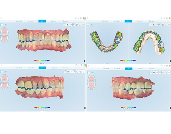
Figure 12
Figure 12: Assessment of the oclussion under Bite and determining the dental relation from the sagittal, frontal, and transverse views. The iTero™ Occlusogram tool is activated to visualize the contacts between the upper and the lower teeth.
Alignment
The iTero™ Occlusogram tool is automatically highlighted under this condition. The patient presents minor crowding in the upper and lower incisors (Figure 13). Orthodontic treatment with Invisalign® aligners would expand the arches in the premolar areas to improve the overall occlusion and facilitate oral hygiene maintenance, with which the patient is struggling.
At this point, we will be focusing on improving the patient’s ability to floss, brush, and use fluoride therapy to prevent current lesions from progressing into cavitations and then in completing the composite restorations and crowns.
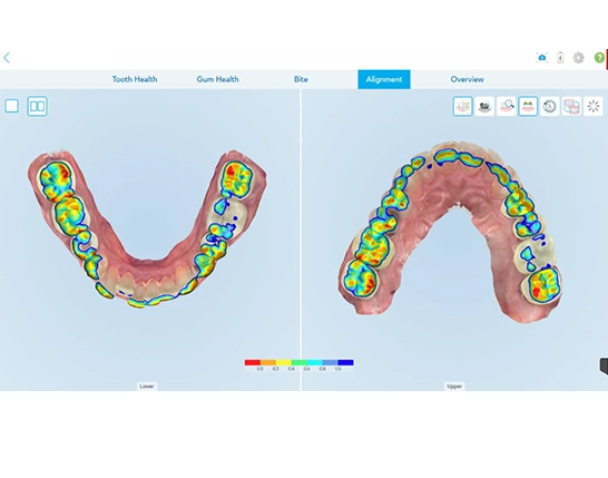
Figure 13
Figure 13: Minor crowding and wear facets are observed in both arches. Orthodontic therapy with Invisalign® aligners could improve the alignment.
The Align™ Oral Health Suite is a helpful new framework on the iTero Element™ Plus Series to conduct your new patient, periodic, or emergency exams. The consolidation of all tools with preset conditions-based views allows for a customized, efficient, and effective exam with increased engagement from prospective or established patients.
It has changed our approach to exams, and we can see how patients have an improved experience with the clarity to understand the treatment plans created for their oral health needs, resulting in increased treatment acceptance and clinical outcomes.

Dr. Joshua Austin runs a busy restorative dentistry practice in San Antonio, Texas.
He also serves as an editorial director and monthly columnist for Dental Economics magazine, where he delves into topics such as dental products and technology.
When he’s not working in his practice or writing, Dr. Austin frequently gives lectures to dental groups across the United States. His areas of expertise also include digital marketing and mental health.
He earned his degree from the University of Texas Health School of Dentistry and spent five years in the Department of Restorative Dentistry after graduation. Since 2018, Dr. Austin has been a member of the Align™ Global Faculty.
The opinions expressed in this publication are those of the author and may not reflect those of Align Technology, Inc. The authors were paid an honorarium by Align Technology, Inc. in connection with this publication.